Home
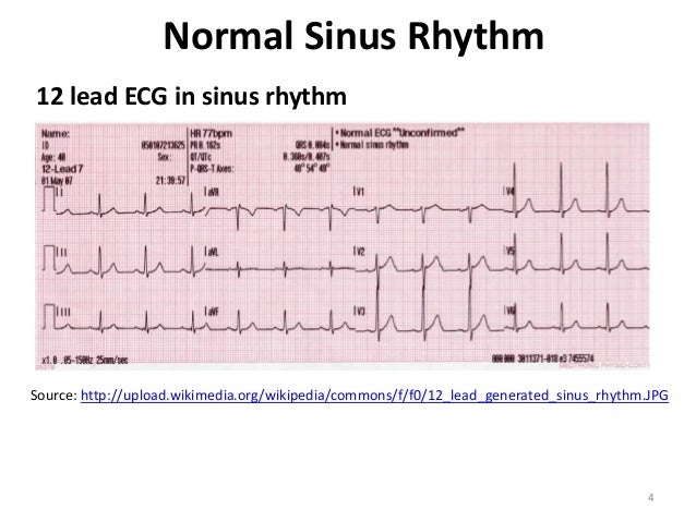
/ Normal Sinus Rhythm Lead Ekg - Ecg Interpretations Ppt Download, Normal sinus rhythm) refers to the normal heart beat originating from the sinoatrial node.
Normal Sinus Rhythm Lead Ekg - Ecg Interpretations Ppt Download, Normal sinus rhythm) refers to the normal heart beat originating from the sinoatrial node.
Normal Sinus Rhythm Lead Ekg - Ecg Interpretations Ppt Download, Normal sinus rhythm) refers to the normal heart beat originating from the sinoatrial node.. Normal sinus rhythm is defined as the rhythm of a healthy heart. It is not considered dangerous, but can escalate to higher level blocks. Anatomical relations of leads in a standard 12 lead electrocardiogram. Rhythm • normal sinus • sinus arrhythmia • sinus arrest. The frequency with which these changes.
During this lecture we will begin by learning how t. Each p wave is followed by a qrs. An axis lying beyond −30° is termed. • a biphid p wave is seen in lead v1. If there is a p wave before each qrs and the p is in the same direction as the qrs, the rhythm can be said to be sinus.
An Artificial Intelligence Enabled Ecg Algorithm For The Identification Of Patients With Atrial Fibrillation During Sinus Rhythm A Retrospective Analysis Of Outcome Prediction The Lancet from els-jbs-prod-cdn.jbs.elsevierhealth.com P waves should be positive and within a normal amplitude (< 2.5mm at lead ii and iii). What is a sinus rhythm in ecg? This condition represents a delay between atrial and ventricular conduction. Sinus rhythm at 75 bpm. Rate <60 = sinus bradycardia. Normal sinus rhythm is defined as the rhythm of a healthy heart. Normal sinus rhythm sinus rhythm is the normal regular rhythm of the heart set by the natural pacemaker of the heart called the sinoatrial node. Normal sinus rhythm the p waves in leads i and ii must be upright (positive) if the rhythm is coming from the sinus node.
Hence, sinus rhythm is the normal rhythm of the heart.
This article deals mainly with ecg features of sinus rhythm. Heart rate will fall between 60 and 100 beats per minute. Sinus rhythm at 75 bpm. Heart rate between 60 bpm and 100 bpm sinus rhythm has a heart rate higher than 60 bpm and lower than 100 bpm. P waves normal for the subject. What is a sinus rhythm in ecg? Normal sinus rhythm sinus rhythm is the normal regular rhythm of the heart set by the natural pacemaker of the heart called the sinoatrial node. Anatomical relations of leads in a standard 12 lead electrocardiogram. Key points from example ecg. The normal range for the cardiac axis is between −30° and 90°. It means the electrical impulse from your sinus node is being properly transmitted. The physiology of the sa node and pacemaker cells in the heart have been discussed previously. This condition represents a delay between atrial and ventricular conduction.
It's description is therefore quite lengthy. Normal sinus rhythm) refers to the normal heart beat originating from the sinoatrial node. P waves normal for the subject. Normal rhythms • sinus tachycardia: This is manifested as an upright p wave in lead ii of the ecg.
Acls Ce Part I Of Iii Ecg Strip Interpretation W Case Scenarios Sup from image.slidesharecdn.com The physiology of the sa node and pacemaker cells in the heart have been discussed previously. Normal sinus rhythm sinus rhythm is the normal regular rhythm of the heart set by the natural pacemaker of the heart called the sinoatrial node. What is a sinus rhythm in ecg? I had an 12 lead ecg results were vent rate 102bpm, pr. Heart rate between 60 bpm and 100 bpm sinus rhythm has a heart rate higher than 60 bpm and lower than 100 bpm. • if the normal ecg is known then interpretation of abnormals becomes easier. Normal p wave morphology and axis (upright in i and ii, inverted in avr) narrow qrs complexes (110 ms wide) Hence, sinus rhythm is the normal rhythm of the heart.
An axis lying beyond −30° is termed.
Normal rhythms • sinus tachycardia: A sinus morphology is an upright p wave in lead ii and biphasic (up and down) p wave in lead v1. Normal duration of less than or equal to 0.11 seconds Heart rate between 60 bpm and 100 bpm sinus rhythm has a heart rate higher than 60 bpm and lower than 100 bpm. What does a good sinus rhythm look like? P waves normal for the subject. I had abnormal ekg where it said normal sinus rhythm, cannot rule out anterior infraction, age undetermined, nonspecific t wave abnormalities no longer evident in inferior leads.then my troponin 1 levels came back normal. Key points from example ecg. Sinus rhythm at 75 bpm. Inferior surface of the heart. The sinoatrial (sa) node is the heart's pacemaker under normal circumstances and the rhythm is referred to as sinus rhythm. The term sinus rhythm is used when the rhythm originates in the sinus node and conducts to the ventricles. What is a sinus rhythm in ecg?
Rate <60 = sinus bradycardia. The frequency with which these changes. Uniform in appearance upright w/ normal shape one preceding each qrs nor more than.10 second o pr interval: Key points from example ecg. The shape of the electrocardiogram (ekg) tracing will exhibit certain key attributes to be considered normal, as discussed below.
Ecg Knowledge Amboss from media-us.amboss.com The sinoatrial (sa) node is the heart's pacemaker under normal circumstances and the rhythm is referred to as sinus rhythm. Normal cardiac impulses start there and are transmitted to the atria and down to the ventricles. This is manifested as an upright p wave in lead ii of the ecg. Normal sinus rhythm sinus rhythm is the normal regular rhythm of the heart set by the natural pacemaker of the heart called the sinoatrial node. This is manifested as an upright p wave in lead ii of the ecg. This encounter displays a p wave of over 200 ms or one square away from the qrs, making it 1st degree heart block. If there is a p wave before each qrs and the p is in the same direction as the qrs, the rhythm can be said to be sinus. Each p wave is followed by a qrs.
This is the currently selected item.
Heart rate between 60 bpm and 100 bpm sinus rhythm has a heart rate higher than 60 bpm and lower than 100 bpm. Rhythm analysis indicates normal sinus rhythm (nsr) with a 1st degree heart block. This condition represents a delay between atrial and ventricular conduction. Normal sinus rhythm is the default cardiac rhythm that represents the normal electrical activity through the heart. Uniform in appearance upright w/ normal shape one preceding each qrs nor more than.10 second o pr interval: What does a good sinus rhythm look like? During this lecture we will begin by learning how t. • a biphid p wave is seen in lead v1. A normal sinus rhythm on an ekg will show an equal distance from r wave to r wave and p wave to p wave. Normal sinus rhythm on an ekg. This encounter displays a p wave of over 200 ms or one square away from the qrs, making it 1st degree heart block. An axis lying beyond −30° is termed. Rhythm • normal sinus • sinus arrhythmia • sinus arrest.
In adults, normal sinus rhythm usually normal sinus rhythm ekg. It is not considered dangerous, but can escalate to higher level blocks.